
We provide mentored training in research and scholarship to individuals during several phases of their educational and professional training to develop independent and sustainable academic careers in basic, clinical, and/or translational research. Trainees include undergraduate and graduate students, medical students, post-doctoral fellows, and surgical residents and fellows in Otolaryngology/Head and Neck Surgery.
Research Training
Research training is supported by several NIH training grants. In particular, the R25 Research Education Grant (which followed our long-standing T32 training grant) from the National Institute of Deafness and Communication Disorders at the NIH supports each resident to conduct mentored research for one year during.
Resident trainees select and conduct research projects based on their interests. Projects can reside within established areas of research in the Department, described above, or they can address new avenues of inquiry via collaborations with mentors outside of the Department.
It is our goal is to provide trainees with the skills they need to develop an independent and sustainable research program in areas relevant to Otolaryngology/Head and Neck Surgery with the ability to:
- Design and execute hypothesis-driven research
- Effectively present findings to the public, in oral and written forms
- Write applications for research funding
Many clinical research trainees earn a certificate or masters degree in clinical research methods and collaborative approaches to address a wide variety of research problems.
Training Sites and Facilities
The Virginia Merrill Bloedel Hearing Research Center and the Health Sciences Building at UW have several laboratories conducting basic science and translational research. Within these sites, there are state-of-the-art imaging centers, surgical suites, tissue culture facilities, histology and microtomy suites, equipment for genetic and molecular analyses, electrophysiological measurements in vivo and in vitro, large-scale drug screening, and detailed auditory and vestibular functional testing.
The Otolaryngology Outcomes Research Group performs clinical research and is based in the UW Comparative Effectiveness, Cost & Outcomes Research Center, an interdisciplinary, multi-school clinical research “laboratory.” These Centers are closely affiliated with the UW School of Public Health and UW Institute of Translational Health.
The Fred Hutchinson Cancer Research Center houses laboratories for basic science and translational research, focusing on head and neck cancer, epithelial tumor biology, and viral biology as it relates to carcinogenesis, immunotherapy, and population studies.
There are innumerable other research facilities where departmental and trainee research is performed. For example, Seattle Children’s Hospital houses research programs on vascular anomalies, pediatric communications disorders, and others. The Department of Electrical Engineering at UW houses an engineering research laboratory studying robotic surgical techniques. The Research & Development (R&D) Program at the VA Puget Sound Health Care Center houses numerous research programs and project, and Harborview Medical Center houses the Harborview Injury Prevention & Research Center and many other research programs.
Visit the Research Resources page for links to these sites and for other helpful information.
Resident Research Highlights
New Tools for Quantification of Dysphagia in Rat Models
Steven Leong, MD
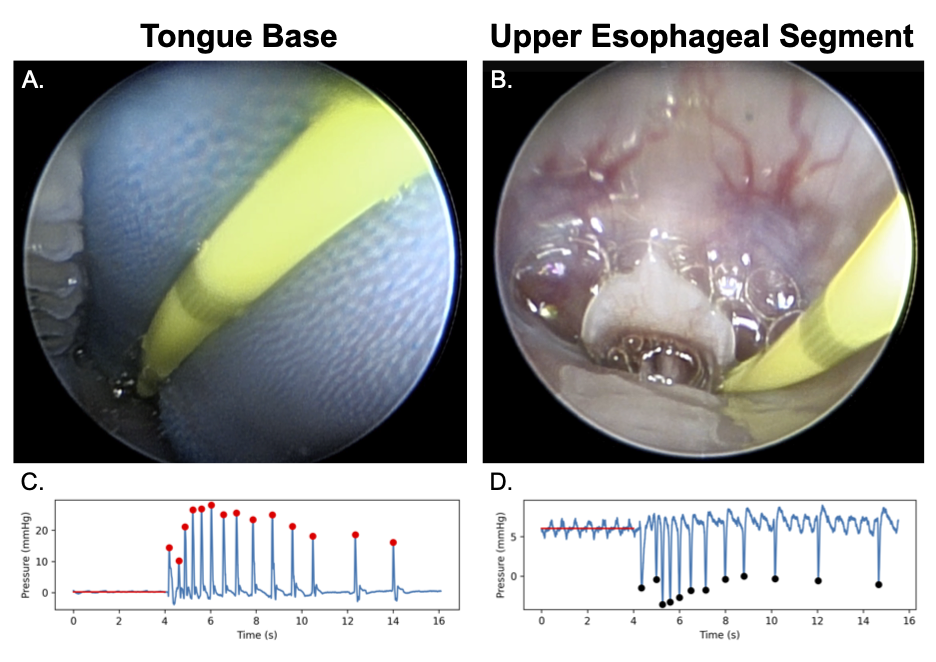
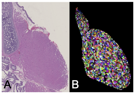
Oropharyngeal dysphagia is commonly observed in aging and neurodegenerative disorders like Alzheimer's disease. Unfortunately, objective quantification of dysphagia remains challenging, and few treatments exist for dysphagia. Under the mentorship of Dr. Neel Bhatt, MD, and Dr. David Perkel, PhD, Dr. Leong has been developing new tools for quantification of dysphagia in various rat models. He has used novel manometric and endoscopic techniques to demonstrate that both aging and Alzheimer's are independently associated with neuromuscular swallow dysfunction. His future work will investigate the structural and neurophysiologic mechanisms of dysphagia in these conditions. Selected laboratory publications are listed here.
A Smartphone Photogrammetry Workflow for 3D Auricular Reconstruction
Rishi Modi, MD
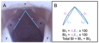
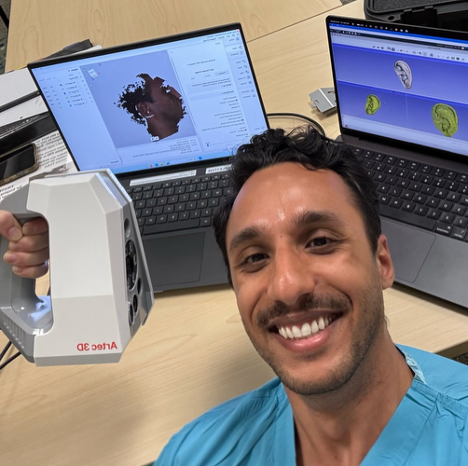
Microtia, anomaly of the external ear, carries significant functional and psychosocial burdens. Repair is commonly done via autologous rib cartilage reconstruction. This technique is extremely complex and relies on 2D photography for surgical planning, and long-term follow up for ideal outcomes. Modi’s R25 research has developed a fully open-source smartphone photogrammetry workflow for 3D auricular reconstruction. His mentors include A. Murat Maga, PhD (Seattle Children’s Research Institute), Kathleen Sie, MD, and Randall Bly, MD (UW Otolaryngology-HNS). Compared with a gold-standard optical scanner, iPhone-derived models were within a clinically accurate threshold. The result is an accessible tool for microtia care, particularly in resource-limited settings.
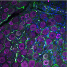
Molecular Subclassification of Mammalian Vestibular Ganglion Neurons Using Single-Cell Transcriptomics
Rahilla Tarfa, MD, PhD
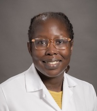
Dr. Tarfa is using single-cell RNA sequencing and complementary experimental techniques to develop a molecular atlas of vestibular ganglion neurons, which transmit information about head movement to the brain. With mentorship from Jennifer Stone and David Raible, and support from an NIH F32 fellowship and American Otological Society funding, she has identified distinct neuronal and glial cell populations and is investigating their roles in vestibular function and neuron-hair cell communication. Her multidisciplinary research combines computational analysis with anatomical and molecular approaches, laying the groundwork for a better understanding of vestibular disorders and, ultimately, targeted strategies for repair and regeneration.